German Rottweilers are
susceptible to hip dysplasia While the Rottweiler puppies from a back yard
breeder may appear healthy, the degradation of the hip joint can take years to
cause noticeable discomfort to the dog. The condition often leads to severe pain
that requires a complete hip replacement. Ethical
Rottweiler breeders will have their breeding stock evaluated by the OFA prior to
any breeding. By eliminating dogs of inferior hip configuration from the
gene pool, the incidence of dysplasia is being greatly reduced in the Rottweiler
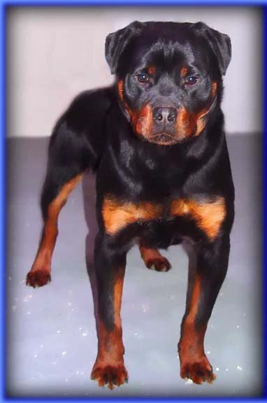
today. Vom Keiser Wappen breeds Rottweilers which have been evaluated as
having a Good or Excellent hip by the OFA.
The OFA is a most prized
resource by identifying those with the condition and keeping a database which
breeders can use freely to evaluate potential breeding combinations.
An introduction to the OFA
John M. Olin was a well-known industrialist,
philanthropist and sportsman. His commitment to the welfare of
dogs was first evidenced in his founding of Nilo Kennels, where he bred, trained
and campaigned outstanding examples of the sporting breeds.
Olin recognized that hip dysplasia was a common
and debilitating orthopedic disease in dogs, and in the fall of 1964 gathered a
group of individuals to discuss means of limiting hip dysplasia. This initial
meeting, which included representatives of the Golden Retriever Club of America,
German Shepherd Dog Club of American, and the veterinary community led to the
organization of the Orthopedic Foundation for Animals (OFA).
The OFA was incorporated as a not-for-profit
corporation by the state of Illinois on July 7, 1966. The original purpose of
the organization was providing radiographic evaluation, database maintenance,
and breeding advice to reduce the incidence of canine hip dysplasia.
Over the past 10 to 15 years the OFA has recognized that a variety of heritable
diseases impact animal health. As scientific advancements enhanced the ability
to diagnose heritable diseases, the OFA has supported development of diagnostic
criteria and databases for a number of genetic diseases in addition to hip
dysplasia.
Hip Dysplasia
Hip Dysplasia is a terrible genetic disease
because of the various degrees of arthritis (also called degenerative joint
disease, arthrosis, osteoarthrosis) it can eventually produce, leading to pain
and debilitation.
The very first step in the development of
arthritis is articular cartilage (the type of cartilage lining the joint) damage
due to the inherited bad biomechanics of an abnormally developed hip joint.
Traumatic articular fracture through the joint surface is another way cartilage
is damaged. With cartilage damage, lots of degradative enzymes are released into
the joint. These enzymes degrade and decrease the synthesis of important
constituent molecules that form hyaline cartilage called proteoglycans. This
causes the cartilage to lose its thickness and elasticity, which are important
in absorbing mechanical loads placed across the joint during movement.
Eventually, more debris and enzymes spill into the joint fluid and destroy
molecules called glycosaminoglycan and hyaluronate which are important
precursors that form the cartilage proteoglycans. The joint's lubrication and
ability to block inflammatory cells are lost and the debris-tainted joint fluid
loses its ability to properly nourish the cartilage through impairment of
nutrient-waste exchange across the joint cartilage cells. The damage then
spreads to the synovial membrane lining the joint capsule and more degradative
enzymes and inflammatory cells stream into the joint. Full thickness loss of
cartilage allows the synovial fluid to contact nerve endings in the subchondral
bone, resulting in pain. In an attempt to stabilize the joint to decrease the
pain, the animal's body produces new bone at the edges of the joint surface,
joint capsule, ligament and muscle attachments (bone spurs). The joint capsule
also eventually thickens and the joint's range of motion decreases.
No one can predict when or even if a dysplastic
dog will start showing clinical signs of lameness due to pain. There are
multiple environmental factors such as caloric intake, level of exercise, and
weather that can affect the severity of clinical signs and phenotypic expression
(radiographic changes). There is no rhyme or reason to the severity of
radiographic changes correlated with the clinical findings. There are a number
of dysplastic dogs with severe arthritis that run, jump, and play as if nothing
is wrong and some dogs with barely any arthritic radiographic changes that are
severely lame.